

San Diego Dental Health Center UCSD
Strategic Panoramic Analysis: Understand the Panoramic Images, learn to recognize artifacts and real pathologies
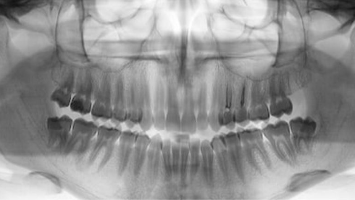
- Radiology
Dentoflow AI summary
This training equips dental professionals with a systematic approach to panoramic radiograph interpretation.
About this training
Content
Panoramic radiographs provide a single-film view of the maxilla, mandible, temporomandibular joints, teeth, and supporting structures. They are frequently used to evaluate unerupted third molars, orthodontic needs, tooth development, developmental abnormalities, trauma, or large lesions. Structures outside the focal trough may appear distorted, magnified, or reduced, so understanding these limitations is essential. The session reviews radiographic physics, common positioning errors, double images, and ghost images. A step-by-step strategic analysis method is demonstrated for reviewing the entire image, followed by clinical cases that illustrate pathology recognition.
Goals
Dental professionals must distinguish normal anatomical landmarks from artifacts or pathology on panoramic images. Systematic review helps avoid overlooking deviations from normal. Panoramic radiography images a broad anatomic region including the mandibular body, ramus, temporomandibular joint, maxillary sinus, and stylohyoid complex, though magnification and geometric distortion can occur when objects lie outside the focal trough.